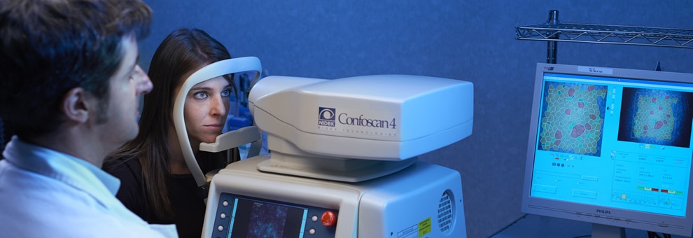
Corneal confocal microscopy (CONFOSCAN)
Corneal confocal microscopy allows performing an “in vivo” analysis of all corneal layers, the anterior part of the eye; in this way it is possible to distinguish individual cells, nerves and other components of the corneal tissue and consequently identify any pathological states.
The patient’s eye is examined without invasive procedures or direct contact. This ensures that any possible discomfort is avoided during the examination.
This examination is indicated for close study of acute and chronic corneal diseases, such as keratoconus, different types of bacterial keratitis and corneal ulcers, and is also advisable for all diseases of the corneal endothelium, especially to determine its integrity before performing cataract surgery.
Want to learn more?


